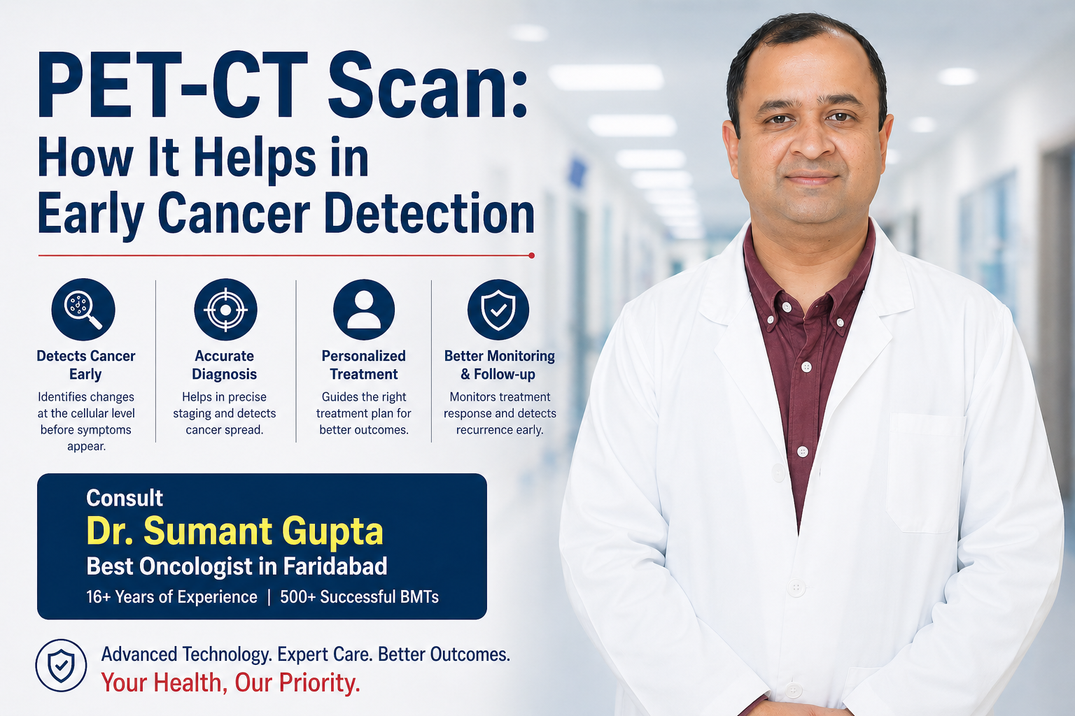
PET-CT Scan: How It Helps in Early Cancer Detection
Oct 30, 2024
Staging describes how far the cancer has spread and guides every major decision: surgery vs. non-surgical treatment, type of chemotherapy, targeted or immunotherapy, and expected outcomes. In lung cancer, doctors use the internationally accepted TNM system—which looks at the Tumor size/extent, Node (lymph node) involvement, and Metastasis (spread to distant organs)—to assign an overall stage from 0 to IV.
In Faridabad, patients typically undergo a streamlined sequence: symptom review → imaging → tissue biopsy → staging work-up → molecular testing. Done correctly, this avoids delays and ensures the right treatment first time.
Common symptoms: persistent cough, coughing blood, chest pain, breathlessness, wheeze, recurrent chest infections, hoarseness, unexplained weight loss, fatigue.
Risk factors: smoking or prior smoking, second-hand smoke, occupational exposures (asbestos, silica, diesel), air pollution, radiation exposure, family history.
Screening: High-risk individuals (e.g., long-term smokers) may be advised Low-Dose CT (LDCT) screening, which can detect cancer earlier than chest X-rays.
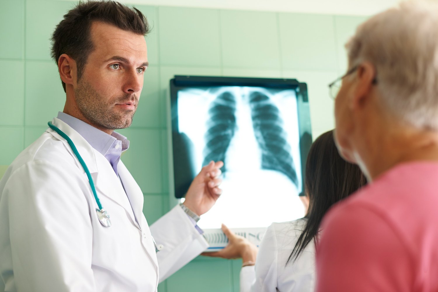
Chest X-ray: often the first look; may miss small or early tumors.
Contrast CT scan (thorax & upper abdomen): defines tumor size, relation to airways, pleura, vessels, and picks up suspicious lymph nodes or liver/adrenal lesions.
PET-CT (FDG): highlights metabolically active areas and improves accuracy of staging by revealing hidden nodal or distant disease.
No lung cancer diagnosis is complete without histopathology (microscopic confirmation). The approach depends on where the lesion is located:
Bronchoscopy (for central lesions): a thin scope is passed into the airways to sample the tumor.
EBUS-TBNA (Endobronchial Ultrasound): samples lymph nodes in the mediastinum with precision.
CT-guided transthoracic needle biopsy (for peripheral nodules).
Thoracoscopy (VATS) or surgical biopsy if needle methods are inconclusive.
Non-Small Cell Lung Cancer (NSCLC) (~85%):
Adenocarcinoma (most common, often peripheral)
Squamous cell carcinoma (often central)
Large cell carcinoma
Small Cell Lung Cancer (SCLC) (~15%): fast-growing, often early spread; typically treated with systemic therapy and radiotherapy.
T (Tumor): size (e.g., ≤3 cm vs >5 cm), invasion into pleura, chest wall, diaphragm, main bronchus, or nearby structures.
N (Nodes): which lymph node stations are involved (same side hilar, mediastinal, opposite side, supraclavicular).
M (Metastasis): spread to the other lung, pleura/pericardium with malignant effusion, bones, liver, adrenal, brain, etc.
Tools used: CT, PET-CT, brain MRI (when indicated), EBUS/mediastinoscopy for nodal confirmation.
Modern lung cancer care relies on molecular profiling of the biopsy:
Driver mutations/fusions: EGFR, ALK, ROS1, BRAF, MET, RET, NTRK, KRAS (e.g., G12C).
PD-L1 expression: helps determine suitability for immunotherapy.
These tests enable targeted therapy or immunotherapy, often improving outcomes and quality of life.
Think of stages as how big the tumor is, which nodes are involved, and whether it has spread elsewhere.
What it means: Very early, abnormal cells confined to the inner lining; no invasion.
Typical management: Surgical removal or ablative techniques; excellent prognosis.
Meaning: Tumor confined to the lung, no lymph nodes involved.
Primary treatment: Surgery (lobectomy/segmentectomy) when fit; sometimes adjuvant (post-op) chemo or targeted therapy depending on size and molecular markers.
Meaning: Larger tumor and/or limited spread to nearby nodes (same side hilar) or chest wall involvement.
Treatment: Surgery plus adjuvant chemotherapy; select cases may receive post-op radiotherapy or targeted therapy based on biomarkers.
Meaning: Mediastinal lymph nodes or locally advanced invasion; often unresectable.
Treatment: Usually concurrent chemoradiation. If no progression after chemoradiation and PD-L1 is suitable, consolidation immunotherapy may follow.
Surgical role: In carefully selected IIIA (after multi-disciplinary evaluation), surgery may be considered.
Meaning: Spread to the other lung, malignant pleural/pericardial effusion, or distant metastases (brain, bone, liver, adrenal, etc.).
Treatment focus: Systemic therapy tailored by molecular profile and PD-L1—targeted therapy, immunotherapy (solo or with chemo), or chemo-immunotherapy combinations; local treatments (stereotactic radiotherapy, surgery) for oligometastatic sites may be considered.
SCLC traditionally uses two practical stages:
Limited-stage: Confined to one hemithorax and regional nodes, potentially treatable with concurrent chemoradiation.
Extensive-stage: Spread beyond a single radiation field; treated with systemic therapy (chemo ± immunotherapy) and palliative radiotherapy as needed.
| Step | What you get | Why it matters |
|---|---|---|
| Consultation | Symptom & risk review | Guides the right tests |
| CT/PET-CT | Tumor & spread map | Core of TNM staging |
| Biopsy/EBUS | Tissue confirmation | Confirms cancer type |
| Histopathology | NSCLC vs SCLC | Decides treatment path |
| Molecular tests | EGFR/ALK/ROS1… PD-L1 | Opens targeted/immunotherapy |
| Multidisciplinary Tumor Board | Combined expert plan | Personalised, stage-wise care |
Q1. Can lung cancer be diagnosed without a biopsy?
No. Imaging can look suspicious, but biopsy is essential to confirm cancer and determine the exact type and gene markers.
Q2. How fast do I need a PET-CT?
Once cancer is suspected or confirmed, staging should be completed promptly to avoid delays in starting the correct treatment.
Q3. If I’m Stage I or II, will I always need chemotherapy after surgery?
Not always. It depends on tumor size, node involvement, and molecular findings. Your team will weigh benefits vs. side-effects.
Q4. I’m Stage IV—do I still have options?
Yes. Many Stage IV patients benefit from targeted drugs or immunotherapy, often with good symptom control and improved survival.
Q5. Do all NSCLC patients need gene testing?
All advanced NSCLC should have comprehensive molecular profiling. Even some early-stage cases can benefit from targeted adjuvant therapy.
Q6. What about brain scans?
A brain MRI is advised in many Stage II–IV cases, adenocarcinoma, or when neurologic symptoms are present.
Q7. I never smoked. Can I still get lung cancer?
Yes. Non-smokers—especially with adenocarcinoma—can carry targetable mutations like EGFR. Testing is crucial.
Q8. How do I choose the right hospital/doctor in Faridabad?
Look for a multidisciplinary team, access to advanced diagnostics (PET-CT, EBUS), molecular testing, and experience with targeted & immunotherapies.
Dr. Sumant Gupta — Best Oncologist in Faridabad and a leading lung cancer specialist in Faridabad — is known for evidence-based, patient-centric cancer care encompassing advanced diagnostics, stage-wise planning, and modern systemic therapies (targeted and immunotherapy).
Book an Appointment Today
Call: +91 981 862 8242
Stage-driven decisions: clear criteria for surgery, chemoradiation, or systemic therapy.
Genomics first: rapid, comprehensive molecular panel and PD-L1 to avoid “trial-and-error.”
Quality radiology & pathology: subtyping accuracy and precise nodal sampling (EBUS, mediastinoscopy).
Tumor board culture: surgeons, medical and radiation oncologists, pulmonologists, radiologists, and pathologists decide together.
Supportive care: nutrition, pulmonary rehab, infection control, pain and symptom management, psychosocial support.
Cough >3 weeks, coughing blood
New/worsening breathlessness or chest pain
Unexplained weight loss or persistent hoarseness
New headaches, bone pain, or neurological symptoms in known lung cancer
Staging (0–IV) tells us how far lung cancer has spread and what treatment fits best.
Biopsy plus molecular testing are non-negotiable in modern care.
A coordinated team improves speed to treatment and outcomes.
In Faridabad, Dr. Sumant Gupta provides the specialist expertise and comprehensive pathway you need—from first CT to final treatment plan.
Oct 30, 2024
Oct 30, 2024
Oct 30, 2024